

IMEMR
EMRO
Volume 7, Issue 2 (2026)
J Clinic Care Skill 2026, 7(2): 51-56 |
Back to browse issues page
Article Type:
Subject:




Ethics code: IR.YUMS.REC.1403.029
History
Received: 2026/01/23 | Accepted: 2026/03/25 | Published: 2026/04/15
Received: 2026/01/23 | Accepted: 2026/03/25 | Published: 2026/04/15
How to cite this article
Ahmadi S, Ariya A, Gorjipour N, Manzouri L. The Relationship Between D-dimer Serum Levels and Diabetic Retinopathy in Type 2 Diabetes Patients. J Clinic Care Skill 2026; 7 (2) :51-56
URL: http://jccs.yums.ac.ir/article-1-496-en.html
URL: http://jccs.yums.ac.ir/article-1-496-en.html
Download citation:
BibTeX | RIS | EndNote | Medlars | ProCite | Reference Manager | RefWorks
Send citation to:


Rights and permissions
BibTeX | RIS | EndNote | Medlars | ProCite | Reference Manager | RefWorks
Send citation to:


1- Department of Internal Medicine, Faculty of Medicine, Yasuj University of Medical Sciences, Yasuj, Iran
2- “Department of Internal Medicine, Faculty of Medicine” and “Social Determinants of Health Research Center”, Yasuj University of Medical Sciences, Yasuj, Iran
2- “Department of Internal Medicine, Faculty of Medicine” and “Social Determinants of Health Research Center”, Yasuj University of Medical Sciences, Yasuj, Iran
Full-Text (HTML) (2 Views)
Introduction
Diabetes mellitus is one of the most significant global health issues and among the most common endocrine disorders [1, 2]. It occurs at all ages and progresses chronically, imposing a substantial economic and social burden on healthcare systems worldwide, particularly in low- and middle-income countries where prevalence is rising most rapidly [3]. Diabetes is categorized into two main types: type 1 diabetes, which is associated with insufficient insulin secretion, and type 2 diabetes (T2D), characterized by insulin resistance and impaired insulin function [4]. These changes lead to metabolic and morphological disturbances in organs such as the kidneys, retina, and blood vessels, affecting nearly all body systems [5, 6]. The global burden of diabetes is escalating at an alarming rate. According to the International Diabetes Federation, approximately 537 million adults were living with diabetes in 2021, and this number is projected to reach 783 million by 2045, with T2D accounting for over 90% of all cases [7].
The complications of T2D are divided into two main categories: microvascular complications (caused by damage to small blood vessels) and macrovascular complications (resulting from damage to larger blood vessels) [8]. Microvascular complications include diabetic retinopathy (DR), diabetic nephropathy, cardiomyopathy, and diabetic neuropathy [9]. DR is one of the most common microvascular complications and a leading cause of blindness among middle-aged and elderly individuals with diabetes [8, 9]. According to the International Diabetes Federation, approximately 9.3% of the global population (463 million people) had diabetes in 2019, and this figure is projected to rise to 10.9% (700 million people) by 2045 [10]. In Iran, estimates indicate that about 11.5% of the population (over 3 million people) are affected by this disease [11]. Among these, DR is not only one of the most common microvascular complications but also a leading cause of preventable blindness among working-age adults, significantly reducing patients’ quality of life and contributing to increased socioeconomic costs [12].
Moreover, the prevalence of DR among diabetic patients is estimated to be approximately 22-27% in the Middle East and North Africa region, emphasizing the critical importance of identifying modifiable risk factors in this population [13, 14]. Vascular complications of diabetes account for nearly 50% of mortality among diabetic patients in developed countries [15]. The underlying pathophysiology of these complications is a complex interplay of metabolic and hemodynamic factors, with chronic hyperglycemia serving as the primary initiator of a cascade of damaging events [16]. Strict glycemic control has been shown in some prospective studies to reduce the incidence and progression of microvascular complications in diabetic patients [17]. However, a significant number of patients continue to develop complications despite optimal glycemic control, suggesting the involvement of additional factors beyond hyperglycemia, including genetic predisposition, oxidative stress, and chronic low-grade inflammation [18]. Chronic hyperglycemia and associated inflammation lead to endothelial damage and impairment of vascular function. Endothelial dysfunction and increased coagulability are among the earliest signs of this process, often preceding clinical manifestations by several years and contributing to a prothrombotic state that exacerbates microvascular occlusion and tissue hypoxia [17, 19]. Therefore, identifying high-risk patients and finding effective strategies for preventing microvascular complications in diabetic patients is of great importance. One of the biomarkers that may be useful in predicting the risk of vascular complications in diabetic patients is D-dimer. D-dimer is a small peptide formed from fibrin degradation in blood clots and serves as an indicator of fibrinolytic activity. Elevated levels of D-dimer signify increased fibrin formation and a tendency toward thrombosis. This marker is routinely used in the diagnosis of vascular thrombosis; however, its elevation can also be observed in other conditions, such as inflammation, cancer, liver disease, pregnancy, and post-surgery [20–22]. Notably, in the context of diabetes, elevated D-dimer levels reflect not only a hypercoagulable state but also an impaired fibrinolytic capacity, which collectively contribute to the pathogenesis of microvascular damage [23].
Diabetic patients with microvascular complications have higher levels of D-dimer than those without such complications. Kiran Shetty et al. state that D-dimer can play a complementary role in the early diagnosis of diabetes complications and the progression of DR [22]. Additionally, Zhuang et al. demonstrate a close correlation between elevated plasma D-dimer levels and peripheral nerve dysfunction in diabetic patients [21]. Elevated plasma D-dimer levels are also identified as having an independent association with inflammation (measured by IL-6) and abnormal lipid profiles in individuals with T2D [16]. Despite these promising findings, the exact mechanistic role of D-dimer in the pathogenesis of DR remains incompletely understood, and studies specifically investigating its association with different stages of DR (non-proliferative vs. proliferative) are limited and occasionally conflicting [24]. Furthermore, most existing studies have been conducted in Western or Asian populations, with a paucity of data from the Middle Eastern context, where genetic, dietary, and lifestyle factors may influence these associations [25].
Considering the importance of predicting and
preventing microvascular complications in diabetic patients and the potential role of D-dimer as a biomarker, this study aimed to investigate the relationship between plasma D-dimer levels and DR in patients with T2D.
Instrument and Methods
This cross-sectional study was conducted in 2024 at the Yasuj Diabetes Clinic and focused on patients with DR among those with T2D who visited the clinic. The sampling method used was either simple random sampling or systematic sampling. The sample size was determined based on the study by Atli et al. [18], with an α of 0.05 and 80% power. An initial sample size of 6 was calculated for each group, and considering a 15% dropout rate, this was increased to 7 per group. To approximate a normal distribution of the parameter, the sample size in each group was set at 30 patients.
Inclusion criteria were complete consent to participate, age between 30 and 80 years, diagnosis of DR by an ophthalmologist, no use of medications with neurotoxic side effects, no history of venous thromboembolism or pulmonary embolism, and no use of anticoagulant or antiplatelet drugs in the past three months. Exclusion criterion included unwillingness to continue participation.
Data were collected through a demographic questionnaire that included information on age, gender, education, living location, microvascular complications, and the duration of diabetes diagnosis.
Written informed consent was obtained from the patient to publish this study and any accompanying images. Measurements of D-dimer levels were obtained through venous sampling conducted by the researcher using a 5mL disposable syringe and collected in disposable tubes containing sodium citrate. Plasma was separated after centrifugation, and D-dimer levels were measured using the ELISA method.
Data analysis was performed using SPSS 25 utilizing independent t-tests for normally distributed continuous parameters according to the Shapiro-Wilk test, or Mann-Whitney tests for non-normally distributed ones. Spearman correlation coefficients and logistic regression analysis were used to assess predictive factors for retinopathy.
Findings
The mean age for the control and retinopathy groups was 51.33±11.01 and 57.60±8.97 years, respectively. There was a statistically significant difference in age between the two groups (p=0.019), with patients in the retinopathy group being significantly older (Table 1).
Table 1. Comparison of the frequency of gender, diabetes medication use, and underlying diseases between the two groups
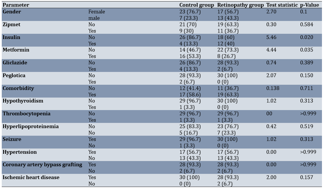
Diabetes duration and D-dimer levels were higher in patients with DR (p=0.001).
Since D-dimer was not normally distributed, it was compared between groups using the Mann–Whitney test and was positively correlated with retinopathy (Spearman’s rho=0.41; p=0.001).
In the multivariable logistic regression model, gender (p=0.018) and D-dimer (p=0.028) were significant independent predictors of retinopathy, whereas age, insulin use, metformin use, and diabetes duration were not significant (Table 2).
Table 2. Determination of predictive factors for retinopathy based on logistic regression
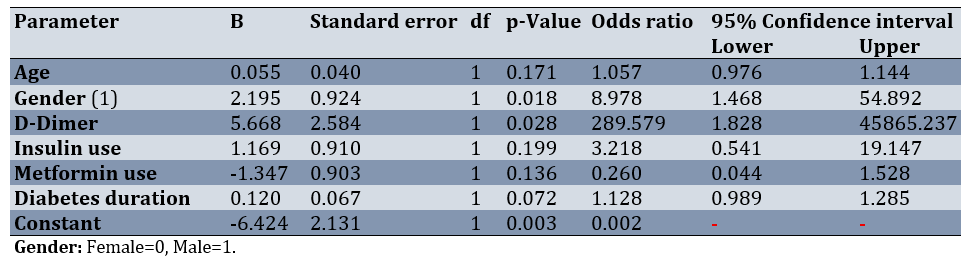
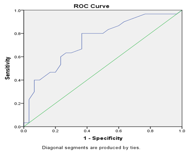
Figure 1. Receiver operating characteristic (ROC) curve for the predictive power of D-dimer levels in diabetic retinopathy complications.
To assess the predictive factors for retinopathy, parameters, including age, gender, duration of diabetes, insulin use status, and metformin use were entered into a logistic regression model.
The receiver operating characteristic (ROC) curve illustrated the predictive power of D-dimer levels for DR complications, with an area under the curve (AUC) of 74% (p<0.05; 95% CI: 0.61-0.87; Figure 1). At a cut-off value of 0.21, the highest sensitivity (80%) and the lowest false positive rate (36%) were observed.
Discussion
This study aimed to investigate the relationship between plasma D-dimer levels and DR in patients with T2D. DR is the most common microvascular complication of diabetes, although its underlying pathology remains incompletely elucidated. Both the duration and severity of hyperglycemia significantly influence the likelihood of developing DR and other diabetes-related microvascular complications [26, 27]. This condition is characterized by impaired retinal perfusion resulting from hyperglycemia-induced microvascular alterations in the retinal vasculature. In its proliferative form, abnormal neovascularization occurs on the optic disc or elsewhere in the retina [28].
This study compared clinical and biochemical parameters between two groups of patients with T2D. Patients with DR were older, had a longer duration of diabetes, and exhibited higher levels of D-dimer than those without DR. Significant differences in medication usage were also observed: the DR group had lower metformin consumption and greater insulin use. Notably, the distribution of gender and the presence of comorbidities did not differ significantly between the two groups.
Zhuang et al. [21] investigated the relationship between plasma D-dimer levels and diabetic peripheral neuropathy (DPN) in patients with T2D. They report a strong association between elevated D-dimer levels and peripheral nerve dysfunction, thereby introducing D-dimer as a promising biomarker for DPN. The results of this study are consistent with ours. Both studies emphasize D-dimer as a marker of vascular diseases in diabetes. Their focus on DPN complements our emphasis on DR, as both conditions involve microvascular damage. This broader perspective strengthens the evidence for using D-dimer as a general predictor of diabetic microangiopathies and further validates your findings.
Cheng et al. [20] conducted a retrospective cohort study to evaluate the role of D-dimer as a predictor of cardiovascular outcomes in patients with T2D. They report that elevated D-dimer levels predict cardiovascular complications and highlight its role as a risk stratification tool. The positive correlation with HbA1c in Cheng et al.'s findings provides additional support for the high D-dimer levels observed in DR, as chronic hyperglycemia is the underlying factor for both microvascular and macrovascular complications. However, Cheng et al. emphasize macrovascular disease, whereas our study focused on microvascular pathology. This distinction illustrates the versatility of D-dimer as a biomarker across a spectrum of vascular complications in diabetes.
Shetty et al. [29] demonstrate that elevated D-dimer levels are significantly associated with advanced stages of DR and high HbA1c levels. The findings of Shetty et al. are directly consistent with ours, as both highlight the importance of D-dimer in the progression of DR and its potential as an early diagnostic marker. The observed association with HbA1c aligns with the pathophysiological link between hyperglycemia and microvascular damage.
In a study conducted by Zhao et al. [30], levels of HbA1c, D-dimer, and fibrinogen are analyzed in 61 diabetic patients at various stages of DR. Higher levels of these markers are observed in proliferative diabetic retinopathy (PDR) compared to non-proliferative diabetic retinopathy (NPDR) and no-DR groups. The combined measurement of these markers improves the early diagnosis of microvascular damage. The findings of Zhao et al. complement our study, further supporting D-dimer as a marker of DR severity. Simó-Servat et al. [31] show that there is a positive association between D-dimer levels and stages of DR, which is consistent with our study.
Collectively, these studies validate D-dimer as a key biomarker for vascular complications in diabetes. While each study focuses on different complications—neuropathy, nephropathy, cardiovascular disease, and retinopathy—they converge on a common theme: Elevated D-dimer levels indicate hypercoagulability and endothelial dysfunction related to the pathophysiology of diabetes. The consistency of the findings across studies justifies the emphasis of our study on D-dimer in DR and demonstrates its broader applicability as a risk stratification tool for diabetic complications. Overall, this study highlights a significant relationship between elevated D-dimer levels and the presence of DR in patients with T2D, suggesting that D-dimer could serve as a potential biomarker for identifying patients at higher risk of DR. The findings underscore the importance of early screening and monitoring of coagulopathy abnormalities in diabetes management.
Conclusion
Diabetic retinopathy is significantly associated with higher D-dimer levels alongside older age, longer diabetes duration, and distinct medication patterns.
Acknowledgments: The authors express their deepest gratitude and appreciation to all the participating patients who collaborated in this study.
Ethical Permissions: This study was approved by the Research Ethics Committee of Yasuj University of Medical Sciences (code number IR.YUMS.REC.1403.029).
Conflicts of Interests: There are no conflicts of interest.
Authors' Contribution: Ahmadi SM (First Author), Introduction Writer/Methodologist/Assistant Researcher/Discussion Writer (45%); Ariya A (Second Author), Introduction Writer/Assistant Researcher (25%); Gorjipour NZ (Third Author), Assistant Researcher (15%); Manzouri L (Fourth Author), Statistical Analyst (15%)
Funding/Support: This study was funded by the respected Research Deputy of Yasuj University of Medical Sciences.
Diabetes mellitus is one of the most significant global health issues and among the most common endocrine disorders [1, 2]. It occurs at all ages and progresses chronically, imposing a substantial economic and social burden on healthcare systems worldwide, particularly in low- and middle-income countries where prevalence is rising most rapidly [3]. Diabetes is categorized into two main types: type 1 diabetes, which is associated with insufficient insulin secretion, and type 2 diabetes (T2D), characterized by insulin resistance and impaired insulin function [4]. These changes lead to metabolic and morphological disturbances in organs such as the kidneys, retina, and blood vessels, affecting nearly all body systems [5, 6]. The global burden of diabetes is escalating at an alarming rate. According to the International Diabetes Federation, approximately 537 million adults were living with diabetes in 2021, and this number is projected to reach 783 million by 2045, with T2D accounting for over 90% of all cases [7].
The complications of T2D are divided into two main categories: microvascular complications (caused by damage to small blood vessels) and macrovascular complications (resulting from damage to larger blood vessels) [8]. Microvascular complications include diabetic retinopathy (DR), diabetic nephropathy, cardiomyopathy, and diabetic neuropathy [9]. DR is one of the most common microvascular complications and a leading cause of blindness among middle-aged and elderly individuals with diabetes [8, 9]. According to the International Diabetes Federation, approximately 9.3% of the global population (463 million people) had diabetes in 2019, and this figure is projected to rise to 10.9% (700 million people) by 2045 [10]. In Iran, estimates indicate that about 11.5% of the population (over 3 million people) are affected by this disease [11]. Among these, DR is not only one of the most common microvascular complications but also a leading cause of preventable blindness among working-age adults, significantly reducing patients’ quality of life and contributing to increased socioeconomic costs [12].
Moreover, the prevalence of DR among diabetic patients is estimated to be approximately 22-27% in the Middle East and North Africa region, emphasizing the critical importance of identifying modifiable risk factors in this population [13, 14]. Vascular complications of diabetes account for nearly 50% of mortality among diabetic patients in developed countries [15]. The underlying pathophysiology of these complications is a complex interplay of metabolic and hemodynamic factors, with chronic hyperglycemia serving as the primary initiator of a cascade of damaging events [16]. Strict glycemic control has been shown in some prospective studies to reduce the incidence and progression of microvascular complications in diabetic patients [17]. However, a significant number of patients continue to develop complications despite optimal glycemic control, suggesting the involvement of additional factors beyond hyperglycemia, including genetic predisposition, oxidative stress, and chronic low-grade inflammation [18]. Chronic hyperglycemia and associated inflammation lead to endothelial damage and impairment of vascular function. Endothelial dysfunction and increased coagulability are among the earliest signs of this process, often preceding clinical manifestations by several years and contributing to a prothrombotic state that exacerbates microvascular occlusion and tissue hypoxia [17, 19]. Therefore, identifying high-risk patients and finding effective strategies for preventing microvascular complications in diabetic patients is of great importance. One of the biomarkers that may be useful in predicting the risk of vascular complications in diabetic patients is D-dimer. D-dimer is a small peptide formed from fibrin degradation in blood clots and serves as an indicator of fibrinolytic activity. Elevated levels of D-dimer signify increased fibrin formation and a tendency toward thrombosis. This marker is routinely used in the diagnosis of vascular thrombosis; however, its elevation can also be observed in other conditions, such as inflammation, cancer, liver disease, pregnancy, and post-surgery [20–22]. Notably, in the context of diabetes, elevated D-dimer levels reflect not only a hypercoagulable state but also an impaired fibrinolytic capacity, which collectively contribute to the pathogenesis of microvascular damage [23].
Diabetic patients with microvascular complications have higher levels of D-dimer than those without such complications. Kiran Shetty et al. state that D-dimer can play a complementary role in the early diagnosis of diabetes complications and the progression of DR [22]. Additionally, Zhuang et al. demonstrate a close correlation between elevated plasma D-dimer levels and peripheral nerve dysfunction in diabetic patients [21]. Elevated plasma D-dimer levels are also identified as having an independent association with inflammation (measured by IL-6) and abnormal lipid profiles in individuals with T2D [16]. Despite these promising findings, the exact mechanistic role of D-dimer in the pathogenesis of DR remains incompletely understood, and studies specifically investigating its association with different stages of DR (non-proliferative vs. proliferative) are limited and occasionally conflicting [24]. Furthermore, most existing studies have been conducted in Western or Asian populations, with a paucity of data from the Middle Eastern context, where genetic, dietary, and lifestyle factors may influence these associations [25].
Considering the importance of predicting and
preventing microvascular complications in diabetic patients and the potential role of D-dimer as a biomarker, this study aimed to investigate the relationship between plasma D-dimer levels and DR in patients with T2D.
Instrument and Methods
This cross-sectional study was conducted in 2024 at the Yasuj Diabetes Clinic and focused on patients with DR among those with T2D who visited the clinic. The sampling method used was either simple random sampling or systematic sampling. The sample size was determined based on the study by Atli et al. [18], with an α of 0.05 and 80% power. An initial sample size of 6 was calculated for each group, and considering a 15% dropout rate, this was increased to 7 per group. To approximate a normal distribution of the parameter, the sample size in each group was set at 30 patients.
Inclusion criteria were complete consent to participate, age between 30 and 80 years, diagnosis of DR by an ophthalmologist, no use of medications with neurotoxic side effects, no history of venous thromboembolism or pulmonary embolism, and no use of anticoagulant or antiplatelet drugs in the past three months. Exclusion criterion included unwillingness to continue participation.
Data were collected through a demographic questionnaire that included information on age, gender, education, living location, microvascular complications, and the duration of diabetes diagnosis.
Written informed consent was obtained from the patient to publish this study and any accompanying images. Measurements of D-dimer levels were obtained through venous sampling conducted by the researcher using a 5mL disposable syringe and collected in disposable tubes containing sodium citrate. Plasma was separated after centrifugation, and D-dimer levels were measured using the ELISA method.
Data analysis was performed using SPSS 25 utilizing independent t-tests for normally distributed continuous parameters according to the Shapiro-Wilk test, or Mann-Whitney tests for non-normally distributed ones. Spearman correlation coefficients and logistic regression analysis were used to assess predictive factors for retinopathy.
Findings
The mean age for the control and retinopathy groups was 51.33±11.01 and 57.60±8.97 years, respectively. There was a statistically significant difference in age between the two groups (p=0.019), with patients in the retinopathy group being significantly older (Table 1).
Table 1. Comparison of the frequency of gender, diabetes medication use, and underlying diseases between the two groups
Diabetes duration and D-dimer levels were higher in patients with DR (p=0.001).
Since D-dimer was not normally distributed, it was compared between groups using the Mann–Whitney test and was positively correlated with retinopathy (Spearman’s rho=0.41; p=0.001).
In the multivariable logistic regression model, gender (p=0.018) and D-dimer (p=0.028) were significant independent predictors of retinopathy, whereas age, insulin use, metformin use, and diabetes duration were not significant (Table 2).
Table 2. Determination of predictive factors for retinopathy based on logistic regression
Figure 1. Receiver operating characteristic (ROC) curve for the predictive power of D-dimer levels in diabetic retinopathy complications.
To assess the predictive factors for retinopathy, parameters, including age, gender, duration of diabetes, insulin use status, and metformin use were entered into a logistic regression model.
The receiver operating characteristic (ROC) curve illustrated the predictive power of D-dimer levels for DR complications, with an area under the curve (AUC) of 74% (p<0.05; 95% CI: 0.61-0.87; Figure 1). At a cut-off value of 0.21, the highest sensitivity (80%) and the lowest false positive rate (36%) were observed.
Discussion
This study aimed to investigate the relationship between plasma D-dimer levels and DR in patients with T2D. DR is the most common microvascular complication of diabetes, although its underlying pathology remains incompletely elucidated. Both the duration and severity of hyperglycemia significantly influence the likelihood of developing DR and other diabetes-related microvascular complications [26, 27]. This condition is characterized by impaired retinal perfusion resulting from hyperglycemia-induced microvascular alterations in the retinal vasculature. In its proliferative form, abnormal neovascularization occurs on the optic disc or elsewhere in the retina [28].
This study compared clinical and biochemical parameters between two groups of patients with T2D. Patients with DR were older, had a longer duration of diabetes, and exhibited higher levels of D-dimer than those without DR. Significant differences in medication usage were also observed: the DR group had lower metformin consumption and greater insulin use. Notably, the distribution of gender and the presence of comorbidities did not differ significantly between the two groups.
Zhuang et al. [21] investigated the relationship between plasma D-dimer levels and diabetic peripheral neuropathy (DPN) in patients with T2D. They report a strong association between elevated D-dimer levels and peripheral nerve dysfunction, thereby introducing D-dimer as a promising biomarker for DPN. The results of this study are consistent with ours. Both studies emphasize D-dimer as a marker of vascular diseases in diabetes. Their focus on DPN complements our emphasis on DR, as both conditions involve microvascular damage. This broader perspective strengthens the evidence for using D-dimer as a general predictor of diabetic microangiopathies and further validates your findings.
Cheng et al. [20] conducted a retrospective cohort study to evaluate the role of D-dimer as a predictor of cardiovascular outcomes in patients with T2D. They report that elevated D-dimer levels predict cardiovascular complications and highlight its role as a risk stratification tool. The positive correlation with HbA1c in Cheng et al.'s findings provides additional support for the high D-dimer levels observed in DR, as chronic hyperglycemia is the underlying factor for both microvascular and macrovascular complications. However, Cheng et al. emphasize macrovascular disease, whereas our study focused on microvascular pathology. This distinction illustrates the versatility of D-dimer as a biomarker across a spectrum of vascular complications in diabetes.
Shetty et al. [29] demonstrate that elevated D-dimer levels are significantly associated with advanced stages of DR and high HbA1c levels. The findings of Shetty et al. are directly consistent with ours, as both highlight the importance of D-dimer in the progression of DR and its potential as an early diagnostic marker. The observed association with HbA1c aligns with the pathophysiological link between hyperglycemia and microvascular damage.
In a study conducted by Zhao et al. [30], levels of HbA1c, D-dimer, and fibrinogen are analyzed in 61 diabetic patients at various stages of DR. Higher levels of these markers are observed in proliferative diabetic retinopathy (PDR) compared to non-proliferative diabetic retinopathy (NPDR) and no-DR groups. The combined measurement of these markers improves the early diagnosis of microvascular damage. The findings of Zhao et al. complement our study, further supporting D-dimer as a marker of DR severity. Simó-Servat et al. [31] show that there is a positive association between D-dimer levels and stages of DR, which is consistent with our study.
Collectively, these studies validate D-dimer as a key biomarker for vascular complications in diabetes. While each study focuses on different complications—neuropathy, nephropathy, cardiovascular disease, and retinopathy—they converge on a common theme: Elevated D-dimer levels indicate hypercoagulability and endothelial dysfunction related to the pathophysiology of diabetes. The consistency of the findings across studies justifies the emphasis of our study on D-dimer in DR and demonstrates its broader applicability as a risk stratification tool for diabetic complications. Overall, this study highlights a significant relationship between elevated D-dimer levels and the presence of DR in patients with T2D, suggesting that D-dimer could serve as a potential biomarker for identifying patients at higher risk of DR. The findings underscore the importance of early screening and monitoring of coagulopathy abnormalities in diabetes management.
Conclusion
Diabetic retinopathy is significantly associated with higher D-dimer levels alongside older age, longer diabetes duration, and distinct medication patterns.
Acknowledgments: The authors express their deepest gratitude and appreciation to all the participating patients who collaborated in this study.
Ethical Permissions: This study was approved by the Research Ethics Committee of Yasuj University of Medical Sciences (code number IR.YUMS.REC.1403.029).
Conflicts of Interests: There are no conflicts of interest.
Authors' Contribution: Ahmadi SM (First Author), Introduction Writer/Methodologist/Assistant Researcher/Discussion Writer (45%); Ariya A (Second Author), Introduction Writer/Assistant Researcher (25%); Gorjipour NZ (Third Author), Assistant Researcher (15%); Manzouri L (Fourth Author), Statistical Analyst (15%)
Funding/Support: This study was funded by the respected Research Deputy of Yasuj University of Medical Sciences.
Keywords:
References
1. Ahmadi S, Gholami L, Malekzadeh J, Hosseni A, Ahmadi S. Comparison of anxiety and depression of type 2 diabetes patients with healthy people during the corona epidemic. ARMAGHAN-E DANESH. 2024;29(3):444-55. [Persian] [Link] [DOI:10.61186/armaghanj.29.3.444]
2. Sherin B, Ramamurthy BD, Sudalaimuthu M, Ganapathy S. Comparison of coagulation profile in type 2 diabetic patients with good glycaemic control and poor glycaemic control. J Clin Diagn Res. 2020;14(7):EC06-8. [Link] [DOI:10.7860/JCDR/2020/44329.13816]
3. Jalilian H, Heydari S, Imani A, Salimi M, Mir N, Najafipour F. Economic burden of type 2 diabetes in Iran: A cost‐of‐illness study. Health Sci Rep. 2023;6(2):e1120. [Link] [DOI:10.1002/hsr2.1120]
4. Shiva A, Maboudi A, Arab S. A review of the complications and oral manifestation of diabetes mellitus. Clin Excell. 2016;5(2):17-27. [Persian] [Link]
5. Seid MA, Akalu Y, Gela YY, Belsti Y, Diress M, Fekadu SA, et al. Microvascular complications and its predictors among type 2 diabetes mellitus patients at Dessie town hospitals, Ethiopia. Diabetol Metab Syndr. 2021;13(1):86. [Link] [DOI:10.1186/s13098-021-00704-w]
6. Bhandarkar NS, Shetty KB, Shetty N, Shetty K, Kiran A, Pindipapanahalli N, et al. Comprehensive analysis of systemic, metabolic, and molecular changes following prospective change to low-carbohydrate diet in adults with type 2 diabetes mellitus in India. Front Nutr. 2024;11:1394298. [Link] [DOI:10.3389/fnut.2024.1394298]
7. International Diabetes Federation. IDF Diabetes Atlas. 7th ed. Brussels: International Diabetes Federation; 2015. [Link]
8. Rasoulinejad S, Akbari A. Vitamin D levels and risk of retinopathy in patients with diabetes mellitus. J Babol Univ Med Sci. 2022;24(1):189-98. [Persian] [Link]
9. Alwin Robert A, Al Dawish MA. Microvascular complications among patients with diabetes: An emerging health problem in Saudi Arabia. Diab Vasc Dis Res. 2019;16(3):227-35. [Link] [DOI:10.1177/1479164118820714]
10. Carmichael J, Fadavi H, Ishibashi F, Shore AC, Tavakoli M. Advances in screening, early diagnosis and accurate staging of diabetic neuropathy. Front Endocrinol. 2021;12:671257. [Link] [DOI:10.3389/fendo.2021.671257]
11. Bahrami Taghanaki H, Mosa Farkhani E, Eftekhari Gol R, Bahrami Taghanaki P, Bokaei S, Taghipour A, et al. Determinants of diabetic complications: A population-based case-control study. Iran J Epidemiol. 2020;16(3):220-9. [Persian] [Link]
12. Gurnani B, Kaur K. Diabetic retinopathy and visual disability. In: The Palgrave encyclopedia of disability. Cham: Springer; 2025. p. 1-13. [Link] [DOI:10.1007/978-3-031-40858-8_520-1]
13. Hazar N, Jokar M, Namavari N, Hosseini S, Rahmanian V. An updated systematic review and Meta-analysis of the prevalence of type 2 diabetes in Iran, 1996-2023. Front Public Health. 2024;12:1322072. [Link] [DOI:10.3389/fpubh.2024.1322072]
14. Samy El Gendy NM, Abdel-Kader AA. Prevalence of selected eye diseases using data harvested from ophthalmic checkup examination of a cohort of two thousand Middle Eastern and North African subjects. J Ophthalmol. 2018;2018(1):8049475. [Link] [DOI:10.1155/2018/8049475]
15. Rasoulinejad SA, Sarreshtehdari N, Mafi AR. The crosstalk between VEGF signaling pathway and long non-coding RNAs in neovascular retinal diseases: Implications for anti-VEGF therapy. Gene Rep. 2022;27:101541. [Link] [DOI:10.1016/j.genrep.2022.101541]
16. Capitão M, Soares R. Angiogenesis and inflammation crosstalk in diabetic retinopathy. J Cell Biochem. 2016;117(11):2443-53. [Link] [DOI:10.1002/jcb.25575]
17. Gerstein HC, Colhoun HM, Dagenais GR, Diaz R, Lakshmanan M, Pais P, et al. Dulaglutide and cardiovascular outcomes in type 2 diabetes (REWIND): A double-blind, randomised placebo-controlled trial. Lancet. 2019;394(10193):121-30. [Link] [DOI:10.1016/S0140-6736(19)31149-3]
18. Atli H, Onalan E, Yakar B, Duzenci D, Dönder E. Predictive value of inflammatory and hematological data in diabetic and non-diabetic retinopathy. Eur Rev Med Pharmacol Sci. 2022;26(1):76-83. [Link]
19. Zheng Y, Ley SH, Hu FB. Global aetiology and epidemiology of type 2 diabetes mellitus and its complications. Nat Rev Endocrinol. 2018;14(2):88-98. [Link] [DOI:10.1038/nrendo.2017.151]
20. Cheng L, Fu Q, Zhou L, Fan Y, Liu F, Fan Y, et al. D-dimer as a predictor of cardiovascular outcomes in patients with diabetes mellitus. BMC Cardiovasc Disord. 2022;22(1):82. [Link] [DOI:10.1186/s12872-022-02531-x]
21. Zhuang L, Yu C, Xu F, Zhao LH, Wang XH, Wang CH, et al. Increased plasma D-dimer levels may be a promising indicator for diabetic peripheral neuropathy in type 2 diabetes. Front Endocrinol. 2022;13:930271. [Link] [DOI:10.3389/fendo.2022.930271]
22. Ceriello A. Diabetes, D-dimer and COVID-19: The possible role of glucose control. Diabetes Metab Syndr. 2020;14(6):1987. [Link] [DOI:10.1016/j.dsx.2020.10.011]
23. Soares AL, De Oliveira Sousa M, Dusse LM, Fernandes APSM, Lasmar MC, Novelli BA, et al. Type 2 diabetes: Assessment of endothelial lesion and fibrinolytic system markers. Blood Coagul Fibrinolysis. 2007;18(5):395-9. [Link] [DOI:10.1097/MBC.0b013e328133f70f]
24. Nguyen TT, Alibrahim E, Islam FA, Klein R, Klein BE, Cotch MF, et al. Inflammatory, hemostatic, and other novel biomarkers for diabetic retinopathy: The multi-ethnic study of atherosclerosis. Diabetes Care. 2009;32(9):1704-9. [Link] [DOI:10.2337/dc09-0102]
25. Mohamed Z, Al-Natour M, Al Rahbi H. Prevalence of diabetic retinopathy among individuals with diabetes in Gulf Cooperation Council countries: A systematic review and meta-analysis. Oman Med J. 2024;39(1):e585. [Link] [DOI:10.5001/omj.2024.77]
26. Matuleviciene-Anängen V, Rosengren A, Svensson AM, Pivodic A, Gudbjörnsdottir S, Wedel H, et al. Glycaemic control and excess risk of major coronary events in persons with type 1 diabetes. Heart. 2017;103(21):1687-95. [Link] [DOI:10.1136/heartjnl-2016-311050]
27. Garg P, Misra S, Yadav S, Singh L. Correlative study of diabetic retinopathy with HbA1c and microalbuminuria. Int J Ophthalmic Res. 2018;4(2):282-6. [Link] [DOI:10.17554/j.issn.2409-5680.2018.04.79]
28. Loho T, Venna V, Setiabudy RD, Sukartini N, Immanuel S, Kumalawati J, et al. Correlation between vitreous advanced glycation end products, and D-dimer with blood HbA1c levels in proliferative diabetic retinopathy. Acta Med Indones. 2018;50(2):132-7. [Link]
29. Shetty K, Lakshmi K, Lobo S, Vivekanad U. Correlation of serum D-dimer levels with stages of diabetic retinopathy and gycosylated hemoglobin levels. Indian J Clin Exp Ophthalmol. 2022;8(2):245-9. [Link] [DOI:10.18231/j.ijceo.2022.049]
30. Zhao H, Zhang LD, Liu LF, Li CQ, Song WL, Pang YY, et al. Blood levels of glycated hemoglobin, D-dimer, and fibrinogen in diabetic retinopathy. Diabetes Metab Syndr Obes. 2021;14:2483-8. [Link] [DOI:10.2147/DMSO.S309068]
31. Simó-Servat O, Planas A, Ciudin A, Simó R, Hernández C. Assessment of advanced glycation end-products as a biomarker of diabetic outcomes. Endocrinol Diabetes Nutr. 2018;65(9):540-5. [Link] [DOI:10.1016/j.endinu.2018.06.003]